Even during MIT’s spring break the halls of the university’s life sciences buildings were still teeming with young students. That’s because on March 29 and 30 scores of students from six area high schools filled the void by the busload to learn about biology and brain science, including from several members of The Picower Institute.
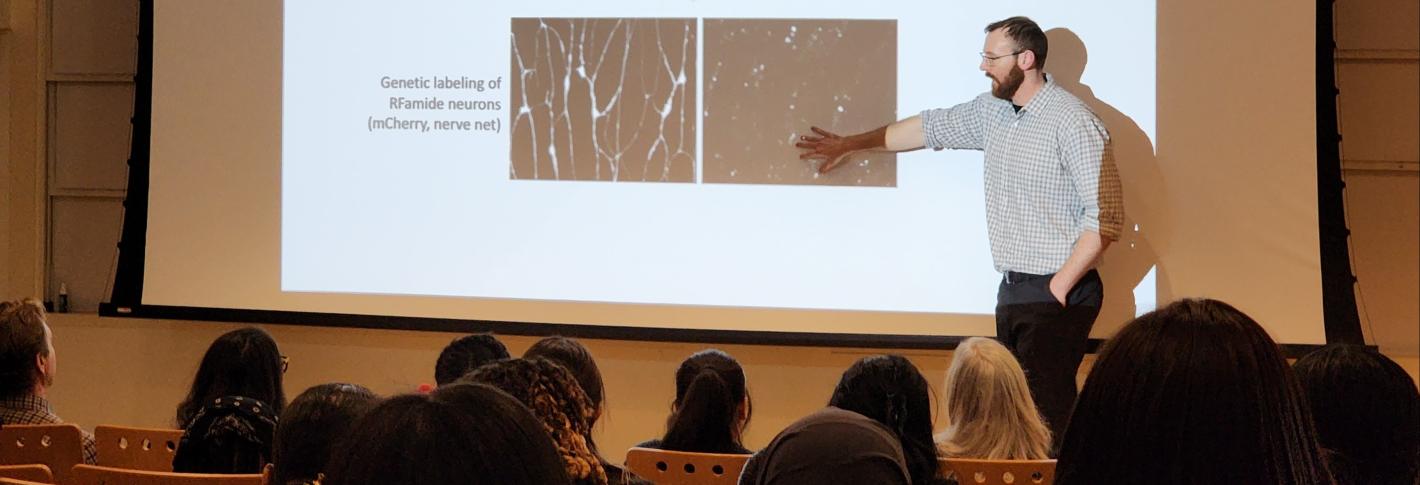
More than 80 students from Everett and Lawrence gathered March 30 to learn about the neuroscience of jellyfish from biology Assistant Professor Brady Weissbourd, a new investigator in The Picower Institute. With vibrant videos and images, Weissbourd explained that the jellyfish nervous system has both amazing differences from humans, like an uncanny ability to regenerate, and important similarities, including the same basic job of generating appropriate behaviors based on their current state and environment.
Above: Assistant Professor Brady Weissbourd describes an experiment with jellyfish neurons to an audience of students from Everett and Lawrence High Schools.
Convenient to study because they are tiny and transparent, jellyfish are also very distant from mammals evolutionarily. So in his lab, which he launched in January, Weissbourd said he plans to learn the secrets jellyfish have to tell about different ways nervous systems have evolved, how their neural organization and activity enables behavior, and how nervous systems can develop and regenerate.
For instance, he’s done experiments where he’s killed all the neurons needed for a jellyfish to execute a particular movement behavior, only to find that within a week they all grow back and the movement ability is restored. Understanding that remarkable resilience could someday help lead to new therapies or technologies, he said.
“New neurons are being born, they are migrating back out and are fully reforming their nervous system entirely from scratch and allowing this behavior to recover,” Weissbourd said. “So a motivation for going out and trying to broadly explore the diversity of biology is that so much of the breakthrough technologies that people have come up with are from understanding some of the amazing capabilities animals can already do.”
On their field trips the visiting teens gained exposure to a wide variety of life science approaches and ideas via demos, tours and talks from numerous labs in the Departments of Biology and Brain and Cognitive Sciences. The tours were coordinated by Mandana Sassanfar, Director of Diversity and Science Outreach.
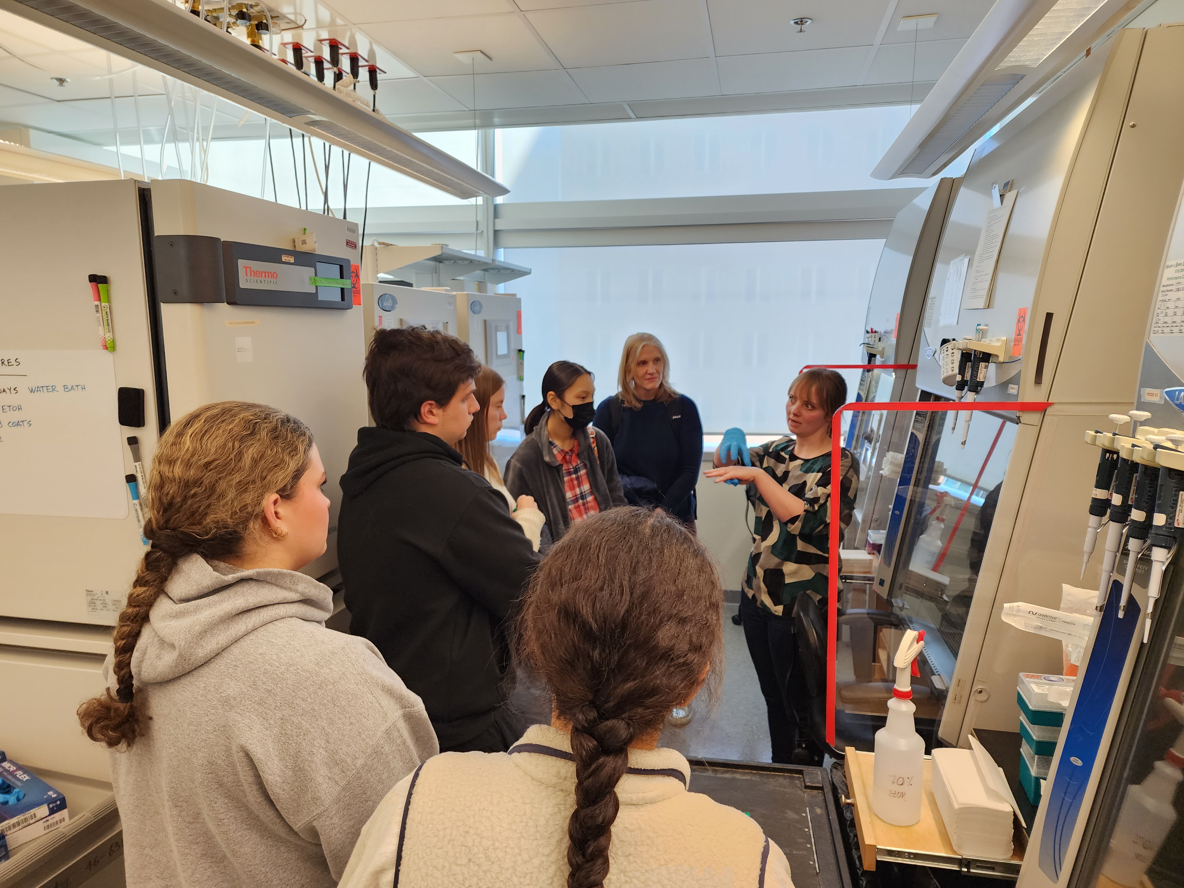
On March 29, for instance, groups of Wellesley students visited with postdoc Matheus Victor and technical associate Áine Ni Scannail to learn how the lab of Picower Professor Li-Huei Tsai cultures human cells to study Alzheimer’s disease. Victor explained that the lab can biopsy an Alzheimer’s patient’s skin cells and reprogram them to become stem cells. He and Scannail showed how those stem cells can then be further manipulated to become neurons or other kinds of brain cells. The resulting human brain cell cultures derived directly from patients can then be used to test how different genes contribute to Alzheimer’s disease pathology and can provide a testbed for whether different drugs could help reduce it.
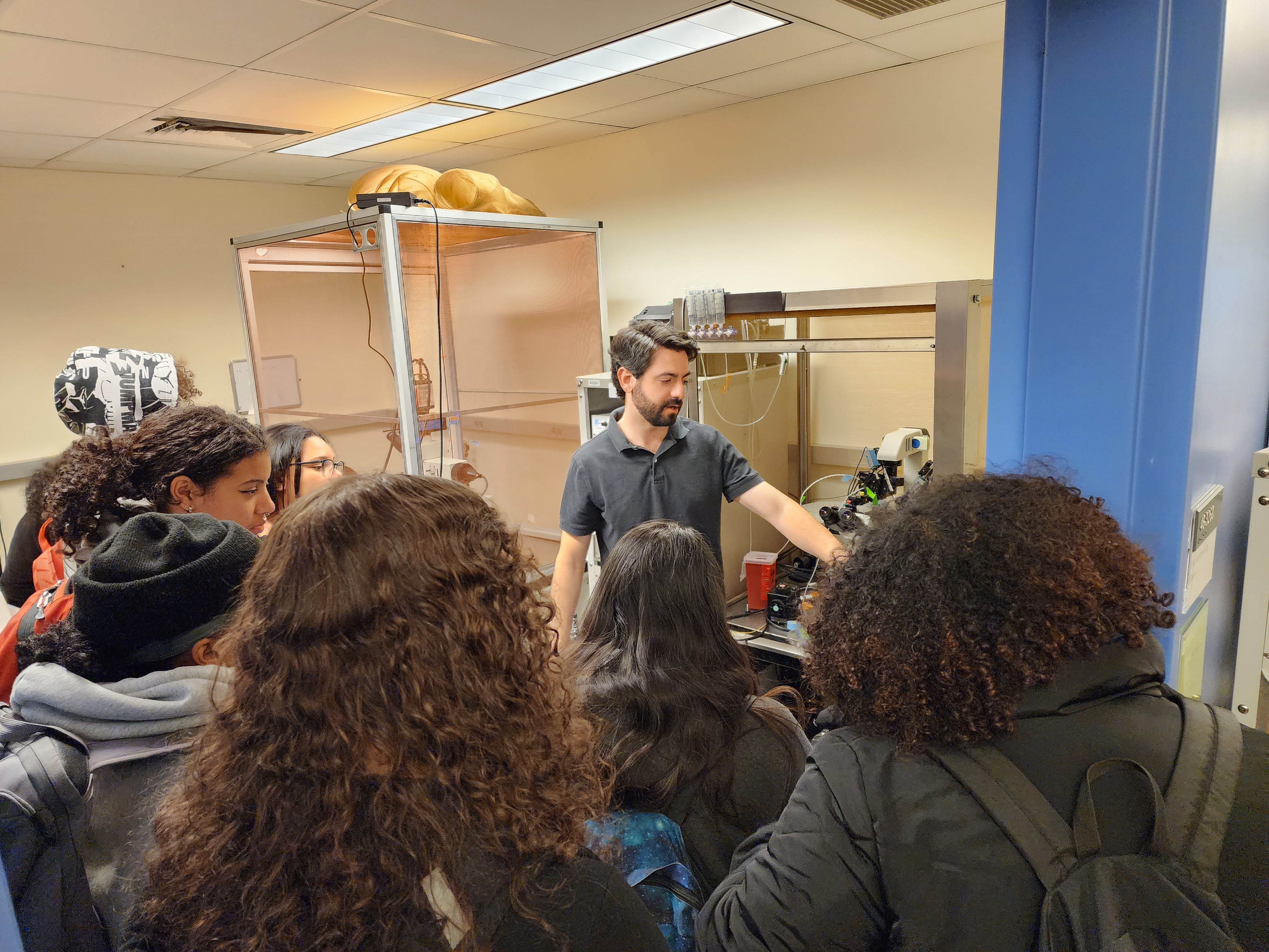
The next day graduate student Andrés Crane led groups of Lawrence High School students through the lab of Menicon Professor Troy Littleton, who uses fruit flies to study fundamentals of how neurons communicate, for instance to control muscle. Crane explained how the researchers can make mutations in the flies to study how those affect the molecules that neurons use to create and operate their connections, or “synapses,” with muscles or other neurons. Crane showed, for instance, how mutations can affect the ability of neurons to transport important molecules, such as the fly version of insulin, from the neuron body down its long axon to synapses at the other end, and how other mutations can affect the strength of a neuron’s electrical signals.
From their visit with Crane the students then went to the lab of Picower Professor Mark Bear, where research scientist David Stoppel showed how he uses mice to study fragile X syndrome, a form of autism. Stoppel demonstrated how he prepares mouse brains so that he can make comparisons about how brain activity differs in neurotypical mice and ones modeling the genetics of fragile X. He showed the high schoolers how brain tissue from the visual cortex of mice modeling the disease shows much greater activity upon electrical stimulation than the tissue from typical mice. That might help explain why people with Fragile X syndrome can experience hypersensitivity to sensory stimulation, he said, and could point to ways to treat that.
“As researchers we have an obligation to use what we learn about the brain to help,” he said.
Many of the visiting high schoolers were seniors approaching graduation later this spring. Perhaps for some, up-close exposure to university research will help shape whether they become part of a next generation of scientists creating new knowledge and ways to help others.


