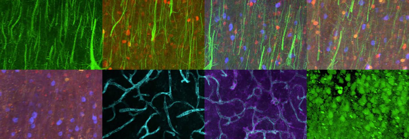
Each kind of nervous system cell can be distinguished by the “fingerprint” of different proteins it expresses. In 2015 Kwanghun Chung's Lab led the development of a new way to classify neurons by labeling and imaging these proteins in cells. Using SWITCH, as they named the method in a paper in Cell, they were able to visualize 22 different proteins inside human brain slices, which could help them tell many different parts of brain anatomy apart.
The ability to vividly resolve all these different molecules not only allows for distinguishing among different cell types, but also to spot molecular differences between cells in healthy and diseased tissue, providing clues to how diseases arise.
The key to SWITCH is that it preserves the structure of the tissue so that it can be labeled and imaged repeatedly, allowing for the full diversity of molecules to come through. It’s as multistep chemical process. Controlling the chemical reactions requires a pair of buffers — solutions of weak acids and bases — that alter the tissue’s environment. One of the buffers, known as SWITCH-Off, halts most chemical reactions in the tissue, while the SWITCH-On buffer allows them to resume.


